

24.05.2017
2 minutes of reading


As part of its research in the field of 2nd generation biofuels, IFPEN is studying the morphology of fungi during the fermentation process. IFPEN’s researchers have developed a new image processing methodology that significantly improves this observation.
Industrial fermentation processes frequently use microscopic fungi, particularly for the production of second-generation fuel where they are employed to produce cellulases, enzymes capable of breaking down biomass. Since the morphology of fungi is a key parameter in the process, its modification to varying degrees in real agitation conditions in reactors tends to detrimentally affect their performance compared to what is expected based on “static” tests conducted in the laboratory.
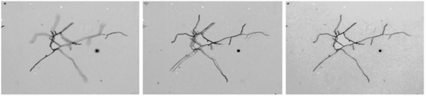
Hence one of the major challenges when it comes to improving enzymatic process performance is being able to accurately observe fungi morphology during the fermentation process. This is difficult because the real morphology of fungi is three-dimensional, i.e., the fungi branches are positioned above and below the principal observation plane. It is also necessary to study a relatively large and representative number of fungi in order to draw conclusions that are statistically significant. Bright-field and short-depth-of-field microscopes can be used to produce high-quality, high-resolution images with an acquisition time compatible with the number of images required for this type of sampling. However, since they can only be used to observe a small sample thickness on the image, it is necessary to produce a series of images at various depths in order to fully observe the fungi.
Our recent research [1] has led to a significant advance in terms of overcoming this challenge, with the contribution of a new algorithm called FACE, or “Fast mean Absolute difference with Confidence propagation for Extended depth of field”. This algorithm makes it possible to create a single merged image that can be exploited and is perfectly clear, from a series of images taken in different planes.
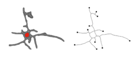
The tool is used in conjunction with a specific skeleton segmentation and calculation approach in order to conduct extensive morphological and topological analyses, with the entire procedure included in a totally automated image processing method, making it possible to analyze a greater number of images than traditionally reported in the literature.
This research was carried out between 2014 and 2016 as part of Nicolas Hardy’s thesis supervised by Catherine Beal (Agro ParisTech), Fadel Ben Chaabane and Frédéric Augier (IFPEN). The image processing algorithms were developed by Denis Guillaume and Maxime Moreaud as part of a project conducted by IFPEN’s Scientific Division.
[1] Advanced digital image analysis method dedicated to the characterization of the morphology of filamentous fungus. N. Hardy, M. Moreaud, G. Denis, F. Augier, A. Nienow, C. Béal, F. Ben Chaabane. Journal of Microscopy, 2017.
>> https://hal.archives-ouvertes.fr/hal-01520793/
Scientific contact : Maxime Moreaud